Axoneme preparation
Bovine sperm
Frozen bovine sperm was obtained from the Utrecht University Veterinary Faculty and was prepared for cryo-EM as previously described2. Briefly, sperm straws were thawed by immersion in a 37 °C water bath for about 30 s. Sperm were then washed twice with Dulbecco’s PBS (Sigma), counted and diluted to concentrations of approximately 0.1–0.2 × 10⁶ cells ml−1 in demembranation buffer (20 mM Tris-HCl pH 7.9, 132 mM sucrose, 24 mM potassium glutamate, 1 mM MgSO4, 1 mM DTT and 0.1% Triton X-100). The suspension was frozen at −20 °C and thawed after 48–96 h. To expose individual DMTs, sperm were then disintegrated by the addition of ATP (Sigma) to a final concentration of 1 mM. Following 10–15-min incubation, about 4 µl of disintegrated sperm was applied to glow-discharged Quantifoil R 2/1 200-mesh holey carbon grids. Using a manual plunger (MPI), grids were blotted opposite the side of cell deposition for 5–6 s then immediately plunged into a liquid ethane–propane mix (37% ethane). Frozen grids were stored under liquid nitrogen until imaging.
Bovine oviduct and porcine brain
Fresh bovine oviducts and porcine brains were sourced from either Trenton Processing Center or the Division of Comparative Medicine at Washington University in St. Louis. No ethical approval or guidance was required, because organs were used from animals killed for other purposes. On receipt, oviduct and brain specimens were carefully opened and brushed, following which they were exposed to an extraction buffer (20 mM Tris pH 7.4, 50 mM NaCl, 1 mM EDTA, 7 mM β-mercaptoethanol, 10 mM CaCl2, 250 mM sucrose and 0.1% CHAPS). The resultant mixture was sieved through a 300-mesh filter to remove tissue debris, followed by 2,000g centrifugation for 5 min to eliminate residual tissue fragments. Subsequently, 13,000g centrifugation for 30 min was performed, and the resulting pellet resuspended in resuspension buffer (RB: 30 mM HEPES pH 7.4, 5 mM MgCl2, 1 mM DTT, 0.5 mM EDTA, 50 mM KCl and Roche protease inhibitor). To enhance purity, we conducted multiple rounds of centrifugation at 2,000g for 2 min and 12,000g for 20 min. Purified cilia were demembranated with 1% NP-40 detergent (Thermo Fisher Scientific) for 1 h at 4 °C, then subsequently centrifuged at 13,000g for 20 min. The resulting pellet was resuspended in 40 μl of RB. To achieve well-separated DMTs, the sample was incubated with 1 mM ATP and 0.02 mg ml−1 subtilisin on ice for 30 min. Samples were applied to Quantifoil R2/1 copper grids mounted in a Vitrobot Mark IV (Thermo Fisher Scientific) operated at 16 °C and 100% humidity. Following blotting for 5 s, grids were plunge-frozen in liquid ethane.
Human oviduct
Human oviduct samples were isolated from whole human uteri from deceased organ donors. The protocol of procurement and processing was reviewed by the Institutional Review Board of Harvard University (protocol no. IRB21-0272), which determined that tissue procurement and processing was not human subject research. No identifying information of deceased organ donors was shared in procurement or processing of tissue.
Human oviduct tissue was provided in PBS (137 mM NaCl, 2.7 mM KCl, 8 mM Na2HPO4 and 2 mM KH2PO4). For purification of human oviduct cilia, individual oviducts were cannulated near the uterotubal junction with a needle of size 18–22G, and gently flushed with 1–3 ml of of PBS using a 10-ml syringe to remove cell debris, vesicles and serous fluid before deciliation. Following a gentle 1–3 ml PBS wash, without removal of the needle, the syringe was filled with 10 ml of deciliation buffer (20 mM HEPES, 10 mM CaCl2, 1 mM EDTA, 50 mM NaCl, 4% sucrose (w/v), 1 mM DTT, 1 mM dibucaine and 1× protease inhibitor cocktail (Sigma, catalogue no. S8830) per 100 ml of deciliation buffer), reconnected to the needle and the mixture flushed through the oviduct into a 50 ml conical tube. Flushing of the oviduct with 10 ml of deciliation buffer was repeated three times for each oviduct before discarding the tissue.
Conical tubes containing eluent were then spun at 900g for 10 min to pellet any large debris dislodged during flushing. Following this low-speed spin, supernatant was then transferred to polycarbonate tubes and spun at 8,000g for 20 min to pellet the human oviduct cilia. The pellet was then resuspended in 100 µl of of RB (30 mM HEPES, 1 mM EGTA, 4 mM MgCl2, 0.1 mM EDTA, 25 mM NaCl, 1× protease inhibitor cocktail (Sigma, catalogue no. S8830) per 100 ml of RB). The resuspended pellet was then examined by negative-stain electron microscopy to determine the degree of cilia isolation. An additional round of pelleting (8,000g for 20 min) was performed, and the pellet resuspended in a volume of about 20–30 µl to achieve a sample with an absorbance reading at 280 nm (A280) of 8–10.
NP-40 at a concentration of 0.5% (v/v) was added to the purified cilia, with rotation at 4 °C for 30 min, to demembranate cilia. The resulting axonemes were then pelleted at 4 °C by centrifuging at 10,000g for 20 min and resuspending in 50 µl of RB to remove any remaining NP-40 detergent. To promote DMT splaying, ATP was then added to the resuspended pellet to a concentration of 2 mM, with rotation at room temperature for 50 min. The sample was then spun at 10,000g for 30 min at 4 °C and resuspended in RB, such that the A280 value of the sample reached 10–12.
Purified human oviduct DMTs, at A280 ranging 10–12 and at volumes of 3 µl, were placed on glow-discharged QF R2/2 grids suspended in a Vitrobot Mark IV at 4 °C and 100% humidity. Following a wait time of 10 s, the grids were blotted for 10–12 s with a force of 10–12 before being plunge-frozen in liquid ethane, then transferred to liquid nitrogen storage.
Porcine oviduct
Oviducts were dissected from intact porcine reproductive tracts shipped overnight, on ice, from Animal Technologies. No ethical approval or guidance was required because organs were used from pigs killed for other purposes. On receipt of the reproductive tract, the oviduct was identified at the tip of the uterine horns and dissected away from the ovary and uterine tissue before being placed in PBS. To purify porcine oviduct cilia, individual oviducts were cannulated near the uterotubal junction with a needle of gauge 18–22G. The oviduct was then gently flushed with 1–3 ml of PBS using a 10-ml syringe to remove cell debris, vesicles and serous fluid before deciliation. Without withdrawing the needle, the syringe was removed and filled with 10 ml of deciliation buffer (20 mM HEPES, 10 mM CaCl2, 1 mM EDTA, 50 mM NaCl, 4% sucrose (w/v), 1 mM DTT, 1 mM dibucaine and 1× protease inhibitor cocktail (Sigma, catalogue no. S8830) per 100 ml of deciliation buffer), reconnected to the needle and the mixture flushed through the oviduct into a 50-ml conical centrifuge tube. Flushing of the oviduct with 10 ml of deciliation buffer was repeated three times for each oviduct before discarding the tissue. Centrifuge tubes containing eluent from flushing of the oviduct were then spun at 900g for 10 min to pellet any large debris dislodged during flushing. Following this low-speed spin, the supernatant was then transferred to polycarbonate tubes and spun at 8,000g for 20 min to pellet the cilia. The pellet was then resuspended in 100 µl of RB (30 mM HEPES, 1 mM EGTA, 4 mM MgCl2, 0.1 mM EDTA, 25 mM NaCl and 1× protease inhibitor cocktail (Sigma, atalogue no. S8830) per 100 ml of RB). Having confirmed the presence of intact cilia in the resuspension by negative-stain electron microscopy, an additional round of pelleting (8,000g for 20 min) was performed. Finally, the pellet was resuspended in about 20–50 µl of RB to achieve a sample with an absorbance reading of 8–15 at A280.
Porcine oviduct cilia at A280 ranging 8–15 were diluted with protein A-conjugated, 10-nm gold fiducials manufactured at an optical density of 10 (OD10) (Cytodiagnostics, catalogue no. AC-10-05-15) to a final gold fiducial concentration corresponding to OD5–10. Next, 3 µl of the mixture was placed on glow-discharged QuantiFoil R2/2 grids suspended in a Vitrobot Mark IV at 4 °C and 100% humidity. The grids were blotted 10 s following sample application for 10–12 s before being plunge-frozen in liquid ethane, then stored in liquid nitrogen before data collection.
Mass spectrometry
Bovine sperm
Proteomics data of bovine sperm were previously reported2 and are available from PRIDE, with accession no. PXD035941.
Bovine oviduct and porcine brain
Demembranated bovine oviduct cilia (BvOv) and porcine brain ventricle cilia (PcBV) were analysed at the Proteomics and Metabolomics Facility at the University of Nebraska-Lincoln. Cilia pellets were resuspended in RB (30 mM HEPES pH 7.4, 5 mM MgCl2, 1 mM DTT, 0.5 mM EDTA and 50 mM KCl) to a final concentration of about 10 mg ml−1. Samples were denatured at 95 °C for 10 min and run approximately 1 cm into an SDS–polyacrylamide gel electrophoresis gel. The gel was fixed for 1 h, washed and stained overnight then destained before either excision of the whole lane (BvOv) or splitting the lane into three fractions (PcBV) for further processing. All gel pieces were washed with water, reduced by the addition of dithiothreitol and alkylated with iodoacetamide before digestion with trypsin overnight at 37 °C. Peptides were dried in a speed vacuum. Digests were redissolved in 2.5% acetonitrile and 0.1% formic acid. Mass spectrometry analyses were carried out using a 2-h gradient on a 0.075 × 250 mm2 C18 Waters CSH column feeding into an Orbitrap Eclipse mass spectrometer run in either OT–OT mode (BvOv) or OT–IT–HCD mode (PcBV).
Samples were analysed using Mascot v.2.7.0 (Matrix Science). Mascot was set up to search a common-contaminants database (cRAP_20150130.fasta, with 125 entries) and either the bovine UniProt database (37,513 sequences, downloaded 23 September 2021) or the porcine UniProt database (49,791 sequences, downloaded 20 June 2022). Mascot was searched with a fragment ion mass tolerance of 0.6 Da and parent ion tolerance of 10 ppm. Deamidation of asparagine and glutamine, oxidation of methionine and carbamidomethylation of cysteine were specified in Mascot as variable modifications.
Human oviduct cilia
Isolated human oviduct cilia were analysed at the Taplin Mass Spectrometry Facility at Harvard Medical School. Cilia were denatured at 95 °C using SDS and run briefly into an SDS–polyacrylamide gel. The gel piece containing ciliary proteins was excised, washed, dehydrated with acetonitrile for 10 min and dried in a speed vacuum. The gel piece was subsequently rehydrated with 50 mM ammonium bicarbonate solution containing 12.5 ng µl−1 trypsin (Promega). After 45 min at 4 °C, the trypsin solution was removed and replaced with 50 mM ammonium bicarbonate solution to cover the gel piece, with incubation at 37 °C overnight. Peptides were later extracted by removal of the ammonium bicarbonate solution, followed by one wash with a solution containing 50% acetonitrile and 1% formic acid. Extracts were then dried in a speed vacuum for around 1 h. Dried peptide samples were reconstituted in 5–10 µl of solvent A (2.5% acetonitrile and 0.1% formic acid), then loaded onto a pre-equilibrated, nanoscale, reverse-phase high-performance liquid chromatography capillary column (100-µm inner diameter × 30-cm (approximate) length) containing 2.6 µm of C18 spherical silica beads. A gradient with increasing concentrations of solvent B (97.5% acetonitrile and 0.1% formic acid) was used to elute peptides with a Famos autosampler (LC Packings). As peptides eluted from the column, they were subjected to electrospray ionization into a Velos Orbitrap Pro ion-trap mass spectrometer (Thermo Fisher Scientific). Tandem mass spectra were acquired and analysed using Sequest (Thermo Fisher Scientific) against a protein database containing normal and reversed versions of all sequences, to determine peptide identities. Data were filtered to a peptide false discovery rate of 1–2%.
SPA cryo-EM data collection
Details of data collection parameters are summarized in Supplementary Table 1.
Bovine sperm
Single-particle analysis cryo-EM data of disintegrated bovine sperm axonemes were collected using either a Talos Arctica operating at 200 kV or a Titan Krios operating at 300 kV (both Thermo Fisher Scientific). Arctica datasets were acquired in super-resolution mode on a K2 Summit direct electron detector (Gatan) with a GIF Quantum energy filter at slit width 20 eV. Krios datasets were acquired on a K3 detector (Gatan) with a BioQuantum energy filter, also with a slit width of 20 eV. Semiautomated data collection was facilitated by SerialEM55, with data quality monitored on the fly using Warp56. Of the 45,431 videos processed, 34,796 were newly collected for this study and were combined with 10,635 from our previously reported dataset2.
Human oviduct cilia
SPA cryo-EM data of disintegrated human oviductal axonemes were collected on a Titan Krios microscope at Harvard Medical School. Videos were recorded on a K3 camera with a BioQuantum energy filter at slit width 20 eV. A 2 × 2 beam tilt pattern with three images per hole was utilized to increase the data collected per stage movement. Each stage position was selected manually to avoid areas with contamination or few DMTs. Images were collected semiautomatically using SerialEM55.
Bovine oviduct cilia and porcine ependymal cilia
SPA cryo-EM data of DMTs from bovine oviduct cilia (4,716 videos) and porcine ependymal cilia (7,051 videos) were collected using Titan Krios microscopes at Case Western Reserve University. Images were collected semiautomatically using SerialEM55.
Cryo-ET data collection
Tilt series were collected on vitrified porcine oviductal cilia using a Titan Krios microscope, operated at a nominal magnification of ×53,000 (corresponding to a pixel size of 1.68 Å) and equipped with K3 camera and a BioQuantum energy filter at slit width 20 eV. SerialEM55 was used for data collection. Targets were selected by identifying cilia in medium-magnification montages. Automatic tilt series were then collected on these targets using a dose-symmetric scheme57, collecting from −54 to +54° with 3° between tilts and targeting a total dose of 110 e/Å2. Data acquisition parameters are summarized in Supplementary Table 1.
SPA cryo-EM data processing
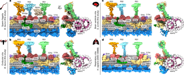
Single-particle data of axonemal DMTs from all cilium types were processed using the same workflow, to ensure consistency between results. All maps reported here represent consensus averages of all nine DMTs, because information about their relative positions is lost during axoneme splaying.
Video frames were drift corrected and dose weighted using patch motion correction in cryoSPARC58. Contrast transfer function (CTF) parameters were estimated using patch CTF estimation in cryoSPARC. DMTs were automatically picked using filament tracer in cryoSPARC. Next, DMT particles were extracted along filament traces using overlapping boxes of 8-nm step size. DMT particles (256-pixel box size, 2× binning) were subjected to two rounds of two-dimensional classification to remove junk and off-centred particles. Good DMT particles then underwent structural refinement using Homogeneous Refinement (New) in cryoSPARC.
Next, these DMT particles and their alignment parameters were exported to FREALIGN v.9.11 and underwent local refinement. In this step we used customized scripts to minimize alignment errors, based on the geometric relationship of neighbouring DMT particles. The particle set with improved alignment parameters was imported back to cryoSPARC for one round of local refinement, followed by tubulin signal subtraction.
For separation of 48-nm repeat from 8-nm particles, we performed three-dimensional classification of tubulin-subtracted DMT particles in Relion 3.1 (ref. 59) using a soft-edged mask covering MIPs near protofilaments A08–A13. A similar strategy was used to further split 48-nm particles into two sets of 96-nm particles, using a soft-edged mask covering an external region near protofilaments A01–A03. The coordinates of 48 and 96-nm DMT particles were imported back to cryoSPARC, re-extracted at 512-pixel box size (no binning) and subjected to one round of local refinement followed by local CTF refinement. This step produced consensus 48 and 96-nm DMT maps. Due to computational constraints, we used a box size of 512 pixels (666 Å) for three-dimensional reconstruction. As a result, we used four different reconstruction boxes whose centres were 24 nm apart (positions 1–4) to cover the 96-nm repeat length (Supplementary Fig. 1).
For improvement of DMT local resolution, we performed focused refinements in cryoSPARC using a set of cylindrical masks as described in ref. 2. These masks divide the DMT into 39 subregions. In regard to external 96-nm features such as RSs and IDAs, we used a similar divide-and-conquer strategy. We first shifted the centre of the nearest reconstruction box (among positions 1–4; Supplementary Fig. 1) to the feature of interest using customized scripts, and then performed three-dimensional classification and focused refinement for the local region (Supplementary Figs. 2–7). In most cases, three-dimensional classification of tubulin-subtracted particles produced better results, and the masks used for three-dimensional classification and focused refinement were adjusted iteratively until no further improvement was observed. For external complexes, a rough total of 20 local regions was refined for each cilium type, with resolution estimates provided in Supplementary Table 2.
To generate a composite map for model building and refinement, we prepared a large rectangular box (600 × 640 × 1,024 pixels) covering the entire length of the 96-nm repeat, by stitching together the two halves of the 96-nm DMT maps. We refer to this rectangular box as the ‘big map’. All reconstructions for local regions were sharpened using deepEMhancer60, which produced a consistent grey level across various maps. Sharpened maps were multiplied by their respective masks and aligned to the big map using the fit in map command in Chimera61. Aligned maps were resampled onto the grid of the big map, and merged using the vop resample and vop maximum commands in Chimera.
Cryo-ET data processing
Videos of ten frames, recorded at each tilt angle, were motion corrected and coarsely aligned into a tilt series of single micrographs using alignframes from IMOD62. Motion-corrected micrographs were manually inspected and removed from the tilt series using etomo if uncorrected drift was observed. Following automatic detection with IMOD, each fiducial position was manually inspected to ensure correct fiducial tracking through the tilt series. The CTF was fit using IMOD’s Ctfplotter, and tomograms were generated in IMOD using back projection.
Within each tomogram, DMTs were manually traced using IMOD’s graphical user interface. Particle positions were then placed every 8 nm along the traced DMTs. Initial translation and angular alignment searches at bin 8 (pixel size 18.24 Å, box 200 pixels) for each particle position were then performed in PEET6 using a 96-nm reconstruction of the T. thermophila DMT (EMD-9023)54 as an initial reference, low-pass filtered to 50 Å. Translational search distances allowed for alignment on the nearest 24-nm repeat. Following initial alignment in PEET, particle positions and Euler angles were then imported to RELION 4.0.1 (ref. 63) for classification, and for additional refinement of alignment and CTF parameters. In RELION, pseudosubtomograms were extracted at each particle position with 8× binning and subjected to a round of local refinement. Subsequently, three-dimensional classification was performed with a cylindrical mask covering the RSs and inner dynein arms (Extended Data Fig. 1e). Classification on density within this cylinder led to separation of 96-nm registers of the DMT, one of which was selected for subsequent reconstruction. Pseudosubtomograms of these particles were then extracted at bins 4, 2 and 1, with local refinement performed before each decrement in bin size. At bin size 1 (pixel size 1.68 Å, box 220 pixels), refinement of CTF parameters and frame alignment was performed to improve the quality of the reconstruction. Because the maximally achieved resolution at bin 1 (8.4 Å, based on the Fourier shell correlation 0.143 criterion) was greater than Nyquist frequency at bin 2 (6.64 Å), subsequent maps of the DMT were aligned at a bin size of 2 or 4.
For reconstruction of a 96-nm map of the porcine oviduct DMT at bin 2 (pixel size 3.36 Å, box 340 pixels), focused refinement was performed on the central 60-nm portion of the DMT. Particles were then shifted along the long axis of the DMT by about 24 nm, followed by a further focused refinement. This was repeated until four overlapping porcine oviduct DMT maps were obtained, shifted by roughly 24 nm and with resolution of 9.2–10.2 Å. A composite 96-nm map was then constructed using vop maximum in ChimeraX64, to record maximum voxel density of the overlapped maps. A map of the 96-nm repeat, including axonemal complexes, was performed at bin 4 (pixel size 6.72 Å, box 340 pixels), with an estimated resolution of 23 Å based on the Fourier shell correlation 0.5 criterion. The resulting map represents a consensus average of all nine DMTs. Note that cryo-ET data were processed independently of SPA data and that the subtomogram average of porcine oviduct DMTs was not used in SPA processing of other axoneme types.
Model building and refinement
Models of the 96-nm axonemal repeat from bovine sperm were built based on available structures of human respiratory axonemes (PDB 8J07)1 and of bovine sperm DMTs (PDB 8OTZ)2. Human proteins were replaced with either predictions from AlphaFold2 (ref. 65) or homology models from SWISS-MODEL66 using the most similar B. taurus sequences from UniProt or NCBI.
Densities unaccounted for by these models were assigned through either sequence- or structure-based approaches. The strategy applied for each newly assigned protein, along with supporting evidence from the literature, is summarized in Supplementary Figs. 8–33. For regions with well-resolved side-chain densities, backbone traces were built either manually with Coot67 or automatically with ModelAngelo68. Either findMySequence69 or ModelAngelo was then used to estimate side-chain probabilities, and to find the best-matching sequence from our bovine sperm proteome. For densities at intermediate resolution (around 5 Å), we applied one of three structure-based approaches, either (1) manual tracing of helices in Coot, followed by querying AlphaFold2 databases using the DALI server70, deepTracerID71 or FoldSeek72; (2) automatic fitting of AlphaFold predictions into segmented density using the colores algorithm73 in the Situs package74, followed by ranking based on cross-correlation scores and manual inspection of top hits75; or (3) using a density-based fold-recognition algorithm based on MOLREP–BALBES, followed by manual inspection of top hits76. To increase confidence in assigning unique proteins to unknown densities, reverse searches were performed using DALI or FoldSeek, and AlphaFold2 predictions of candidate proteins as queries. If multiple proteins could fit equally well, these alternatives are noted in Supplementary Figs. 8–33.
AlphaFold2 predictions of newly identified proteins or protein subcomplexes were fit into the density maps using ChimeraX64, followed by manual adjustment in Coot and molecular dynamics flexible fitting with Namdinator77. Individual PDB files were merged and given unique chain IDs in ChimeraX, then real-space refined in Phenix78 using a non-bonded weight of 500. Due to the size of the model, the 96-nm repeat was split into two halves, each refined independently. Model statistics are summarized in Supplementary Table 5.
Reporting summary
Further information on research design is available in the Nature Portfolio Reporting Summary linked to this article.


