Patients and samples
Ten patients with melanoma, one with breast cancer and one with skin squamous cell carcinoma were enrolled in this study, and samples were used to establish TILs and matched cancer cell lines (cohort A; Supplementary Table 1). Participants had undergone surgical resection at Yamanashi University Hospital, Chiba University Hospital, Shinshu University Hospitals or Saitama Medical University International Medical Center. The tumour tissues were processed as previously described60. In brief, surgically resected samples were enzymatically digested with 0.1% collagenase, 0.01% hyaluronidase and 30 U ml–1 deoxyribonuclease (Sigma-Aldrich) in RPMI1640 (Thermo Fisher Scientific) at room temperature. The digested tumour cells were subjected to filtration and density-gradient separation before use. Peripheral blood mononuclear cells were obtained from donated blood and through Ficoll–Uropoline density-gradient centrifugation. All participants provided written informed consent.
In addition to abovementioned tumour samples, FFPE tissue samples were obtained from three other groups of participants for mtDNA mutation analyses. Cohort B comprised 95 patients with melanoma treated with anti-PD-1 monoclonal antibody and/or anti-CTLA-4 monoclonal antibody as first-line therapy. FFPE tissue samples were obtained between 2014 and 2020 from Yamanashi University Hospital, Chiba University Hospital, Shinshu University Hospital and Saitama Medical University International Medical Center from 2014 to 2020 (Extended Data Table 1). Cohort C1 comprised 86 patients with NSCLC treated with anti-PD-1 monoclonal antibody monotherapy as first-line treatment. FFPE tissue samples were obtained between 2017 and 2021 from Chiba University Hospital, Okayama University Hospital and Kindai University Hospital (Extended Data Table 1). Cohort C2 comprised 56 patients with NSCLC treated with platinum-doublet cytotoxic chemotherapies without any ICIs as first-line treatment. FFPE tissue samples were obtained between 2011 and 2012 from Chiba University Hospital, Okayama University Hospital and Kindai University Hospitals (Supplementary Table 3). Clinical information on the participants was obtained from their medical records. Informed consent was obtained by the patient opting out on the website of our institutions.
The protocol for this study was approved by the appropriate institutional review boards and ethics committees of Yamanashi University Hospital, Chiba University Hospital, Shinshu University Hospital, Okayama University Hospital, Kindai University Hospital and Saitama Medical University International Medical Center. This study was conducted in accordance with the principles of the Declaration of Helsinki.
Sequencing of total mtDNA from clinical samples
Sequencing of total mtDNA from TILs with matched LCL cells, peripheral blood and/or cancer cells from cohort A was performed. mtDNA was isolated from these cells using a QIAmp DNA Mini kit (Qiagen). We also collected FFPE tumour samples from 95 patients with melanoma (cohort B) and 142 patients with NSCLC (cohorts C1 and C2) and matched normal tissue samples from 45 patients for total mtDNA sequencing. Areas with high tumour content were marked in FFPE samples, and mtDNA from these areas was isolated using a GeneRead DNA FFPE kit (Qiagen). A Precision ID mtDNA Whole Genome Panel (Thermo Fisher Scientific) was used to amplify the entire mtDNA sequence using 81 primer pairs, followed by library construction and sequencing on an Ion Torrent Proton sequencer (Thermo Fisher Scientific). After adapter trimming using BaseCaller (with parameter settings –trim-qual-cutoff 15 –barcode-filter-minreads 10 –phasing-residual-filter=2.0 –max-phasing-levels 2 –num-unfiltered 1000 –barcode-filter-postpone 1), reads were mapped to the Revised Cambridge Reference Sequence mitochondrial sequence using TMAP (v.5.12.3; -i bam -v -Y -u –prefix-exclude 5–o 2 stage1 map4) to generate binary alignment maps for subsequent analyses. For each sample, variants were called using Mutect2 in the Genome Analysis Toolkit (v.4.1.8) under mitochondrial mode and with the read filter marked as duplicate disabled. The detected variants were annotated using GRCh38.p14 as the reference by SnpEff (v.5.1d)61 and subjected to additional candidate validation by EAGLE (v.1.1.1)62. Allele frequencies computed by EAGLE (–hetbias=0 and –omega=1e-6) were used as the basis for subsequent variant analyses.
After pooling the EAGLE results from all samples, we used z scores determined from the variant allele frequency (VAF) per position as a metric to evaluate potential variants. Alterations with z scores > 3, VAF > 0.2 and read depths exceeding 100 were selected to minimize false positives from sequencing errors. In cohorts B, C1 and C2, variants with VAF > 0.85 were initially considered polymorphisms and excluded from final reporting under the assumption that somatic variants would not exhibit high VAFs owing to the presence of a substantial fraction of non-cancerous cells in the tumour tissue in the specimens collected. Haplogrep (v.2.4.0; Kulcyznski classification mode with 17_FU1 phylotree, 10 top hits, extended final output and heteroplasmy levels set to 0.85)63 was used to characterize potential haplogroups of individual cases and to identify possible unlabelled SNPs from our frequency-based selection criteria. Variants labelled as ‘hotspot’ and ‘local private variant’ were regarded as polymorphic variants; filtered variants were visually inspected to exclude probable sequencing errors at the termini of PCR amplicons or in homopolymer sequence stretches. For performance assessment, we compared the presence of true variants, confirmed through a paired analysis of matched tumour and normal tissue samples (n = 45), with variants that were called only from tumour samples. Using these criteria, true variants were called with a false-positive rate of 0% and a false-negative rate of 12.2%.
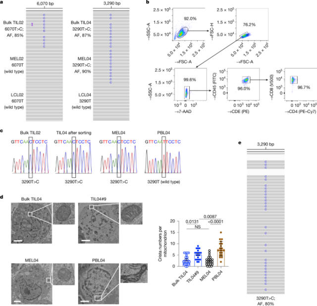
In cohort A, if a sample had LCL, PBL, TIL and/or cancer cell sequencing data, the results were compared with LCLs or PBLs as controls for somatic calling; mutations in TILs or cancer cells were not considered somatic if they existed in the matched LCLs. Using this criterion, ten mtDNA mutations were confirmed in TILs and cancer cells (Supplementary Tables 1 and 2). In cohorts B, C1 and C2, we identified 158 variants, including 48 frameshift and 110 substitution variants, from 237 FFPE tumour samples (Supplementary Table 4). For each sample, the overall mtDNA variant status was classified as truncating, missense, silent, tRNA, rRNA, D-loop or intergenic sites. For survival analysis, samples were divided into mtDNA-mutation-positive if they presented truncating, missense, tRNA or rRNA variants (melanoma, 32.6%; NSCLC (C1), 51.2%; NSCLC (C2), 35.7%) and mtDNA-mutations-negative if they had a D-loop or an intergenic site or had silent or no variants.
Cell lines and culture conditions
To establish cancer cell lines, 1 × 10⁷ digested tumour cells were cultured in RPMI1640 medium containing 10% FBS (Cytiva), 1% penicillin–streptomycin (PS) and 1% amphotericin B (Thermo Fisher Scientific). Tumour cells were passaged at approximately 80–90% confluence and used when free of fibroblasts and proliferating beyond the tenth passage. To establish and expand cultured TILs, tumour digests were incubated in RPMI1640 medium supplemented with 10% human AB serum, 1% PS and recombinant human interleukin-2 (rhIL-2: 6,000 IU ml–1, PeproTech) in a humidified 37 °C incubator with 5% CO2. Half of the medium was aspirated from the wells and replaced with fresh complete medium and rhIL-2 every 2–3 days.
The MC-38 cell line (mouse colon adenocarcinoma) was purchased from Kerafast, and the B16F10 (mouse melanoma), MCF7 (human breast cancer), MDA-MB-231 (human breast cancer) and Jurkat (human T cell leukaemia) cell lines were purchased from the American Type Culture Collection (ATCC). The LLC/P29 and LLC/A11 (mouse lung carcinoma) cell lines were established from mouse Lewis Lung carcinomas as previously described17,64,65. MCF7, MDA-MB-231, MC-38, LLC/P29 and LLC/A11 cells were maintained in Dulbecco’s modified Eagle’s medium (DMEM; Thermo Fisher Scientific), and Jurkat cells were cultured in RPMI1640 medium containing 10% FBS and 1% PS in a humidified 37 °C incubator with 5% CO2. mtDNA-deficient Jurkat (Jurkat/Rho0) cells were generated by culturing Jurkat cells in the presence of 200 ng ml–1 ethidium bromide for 6 weeks and then maintained in RPMI1640 medium containing 10% FBS, 1% PS, 100 μg ml–1 sodium pyruvate and 50 μg ml–1 uridine.
All cell lines were used after confirming that they were mycoplasma-free, which was assessed using a PCR Mycoplasma Detection kit (Takara) according to the manufacturer’s instructions.
Transmission electron microscopy
For pre-fixation, cell specimens were immersed in 0.1 M PBS, pH 7.4, containing 2% glutaraldehyde and 2% paraformaldehyde for 16–18 h. Post-fixation was performed with 2% osmium tetroxide for 1.5 h. After washing with PBS, the specimens were dehydrated in a graded ethanol series and embedded in a low-viscosity resin (Spurr resin, Polysciences). Subsequently, 80-nm-thick sections were prepared using an ultramicrotome (EM-UC7; Leica) and stained with uranyl acetate and lead citrate. The specimens were observed using a transmission electron microscope (H-7650, Hitachi). We counted and quantified the number of cristae per mitochondrion.
Constructs, virus production and transfection
pcDNA3-TfR-OVA (Addgene, 64600)66, pBABE-puro (1764)67 and pVL-MitoDsRed (44386)68 were purchased from Addgene. The TfR-OVA cDNA was cloned into a pBABE-puro vector using In-Fusion Snap Assembly master mix (Takara Bio) according to the manufacturer’s instructions. The resulting pBABE-puro-TfR-OVA vector was transfected with a pVSV-G vector (Takara Bio) into packaging cells using Lipofectamine 3000 reagent (Thermo Fisher Scientific). The pVL-MitoDsRed vector was transfected with packaging plasmids (pMDLg/pRRE, Addgene, 12251 (ref. 69); pRSV-Rev, Addgene, 12253 (ref. 69); and pMD2.G, Addgene, 12259) into packaging cells using Lipofectamine 3000 reagent. After 48 h, the supernatants were concentrated and transduced into cell lines MEL02, MEL04, MELc03, MCF7, MDA-MB-231, B16F10, LLC/P29 and LLC/A11. The transfected cell lines were named MEL02-MitoDsRed, MEL04-MitoDsRed, MELc03-MitoDsRed, MCF7-MitoDsRed, MDA-MB-231-MitoDsRed, B16F10-OVA, LLC/P29-MitoDsRed, LLC/A11-MitoDsRed, respectively. LLC/P29-OVA-MitoDsRed and LLC/A11-OVA-MitoDsRed cell lines were also generated.
Detection of mtDNA
Capillary sequencing and several primers were used to check the status of mtDNA in cell lines and TILs (Supplementary Table 5). For single-cell sequencing, PrimeSTAR GXL DNA Polymerase (Takara) and the primers were prepared in 96-well plates for sequencing before cell sorting. CD3+CD45+ T cells were sorted from TILs at the single-cell level into 96-well plates using a cell sorter (FACSMelody; BD Biosciences). Oligonucleotides were amplified by PCR and sequenced (Eurofins Genomics).
Imaging of mitochondrial transfer
TIL04#9 and MEL04 cells were labelled with MitoTracker Green (Thermo Fisher Scientific) and MitoDsRed, respectively. After 24 h, 2 × 10⁵ MEL04-MitoDsRed cells were cocultured with labelled 1 × 10⁶ TIL04#9 cells in a 35-mm glass-bottom culture dish for 2 days and observed under a Leica TSC SP8 confocal laser microscope (Leica Microsystems) without fixation. TIL04#9 cells were labelled with a BV421-conjugated monoclonal antibody specific for CD45 (clone HI100, BioLegend).
For time-lapse imaging, 2 × 10⁵ MEL04-MitoDsRed cells were seeded onto a 35-mm glass-bottom culture dish 24 h before coculture and allowed to adhere. The following day, coculture with 1 × 10⁶ TIL04#9 cells labelled with MitoTracker Green was initiated. Twenty-four hours later, we began capturing images every 30 min using a digital holographic microscope (3D Cell Explorer CX-A, Nanolive). The images were analysed using Fiji software (https://imagej.net/software/fiji).
Quantification of mitochondrial transfer
To quantify the transfer of mitochondria from cancer cells to TILs, MEL04-MitoDsRed cells were cocultured with TIL04#9 cells for 1, 2, 3 or 14 days with or without 10 mM NAC (Cayman Chemical), 20 nM USP30 inhibitor (CMPD-39; MedChemExpress) or siRNAs for USP30 transfection (siUSP30-1, CAAAAGGUCAUCUGAGGUAAGGCTA; siUSP30-2, CGUCAGAUAUAAAGUCAUGAAGAAC; Integrated DNA Technologies). When using siRNAs, we prepared siRNA-transfected cancer cells every 5 days and cocultured them with TILs, replacing the old, transfected cancer cells. siRNA transfection of cancer cells was performed using Lipofectamine RNAiMAX reagent (Thermo Fisher Scientific) according to the manufacturer’s protocol. To quantify mitochondrial transfer, MEL02-MitoDsRed or MEL04-MitoDsRed cells were cocultured with TILs (TIL02, TIL04#9 or TILc04) for 2 days in the presence of 1:100 anti-MHC-I monoclonal antibody (anti-human HLA-ABC monoclonal antibody, clone W6/32, Thermo Fisher Scientific), 350 nM TNT inhibitor (cytochalasin B; Wako), 1 μM small EV release inhibitor (GW4869; Cayman Chemical) and/or 5 μM microEV release inhibitor (Y-27632; MedChemExpress)30 with or without cell culture inserts to avoid direct cell–cell contact (3 or 0.4 μm pore; Nunc Polycarbonate Cell Culture Inserts in Multi-Well Plates, Thermo Fisher Scientific). Subsequently, TILs were analysed by flow cytometry.
To quantify the transfer of mitochondria from T cells to cancer cells, we used OT-1 and PhaMexcised mice expressing mitochondrial-specific fluorescence (mito-Dendra2), which were obtained from the Jackson Laboratory (Supplementary Fig. 2). OVA-overexpressing LLC/A11 cells were cocultured with or without CD8+ T cells from PhaMexcised OT-1 mice for 3 days. Afterwards, floating T cells were discarded and LLC/A11 cells were repeatedly washed with PBS. These cells were subsequently analysed by flow cytometry as CD45– cells.
Quantification of mitochondria in T cells
To quantify in situ mitochondria in T cells, TIL04#9 cells were labelled with MitoTracker Green for 24 h and then cocultured with MEL04 cells with or without 10 mM NAC for 3 days. To inhibit mitophagy, 100 nM of a mitophagy inhibitor (bafilomycin A1; Adipogen Life Sciences) was added every 12 h for 3 days. Subsequently, the TILs were analysed by flow cytometry.
Quantification of mitochondria in cancer cells
OVA-overexpressing LLC/A11 cells or MEL04 cells were labelled with MitoTracker Green. After 24 h, labelled OVA-overexpressing LLC/A11 cells or MEL04 cells were cocultured with or without CD8+ T cells from OT-1 mice or TIL04#9 cells for 3 days, respectively. Floating T cells were then discarded and OVA-overexpressing LLC/A11 cells or MEL04 cells were repeatedly washed with PBS. These cells were then analysed by flow cytometry as CD45– cells.
Gene expression analysis
TIL04#9 cells were cocultured with MEL04-MitoDsRed cells for 14 days, and DsRed– cells and DsRed+CD3+ T cells were sorted. Total RNA was then extracted using a RNeasy Plus Mini kit (Qiagen) and 100 ng of total RNA was reverse-transcribed into cDNA using Prime-Script RT master mix (Takara). Real-time PCR was performed using PowerUp SYBR Green master mix according to the manufacturer’s instructions. For each sample, the ΔCt for BNIP3, ATF4, IL6, CXCL8 and IL1B versus ACTB (used as an internal control) was calculated as ΔCt = Ct (BNIP3, ATF4, IL6, CXCL8 or IL1B) – Ct (ACTB). The ΔΔCt for DsRed– cells and DsRed+TIL04#9 cells versus TIL04#9 cells was calculated as ΔΔCt = ΔCt (DsRed– or DsRed+TIL04#9 cells) – ΔCt (TIL04#9 cells). The primers are listed in Supplementary Table 5.
Evaluation for mitophagy
TIL04#9 cells were labelled with MitoTracker Green. Twenty-four hours later, TIL04#9 cells were cocultured with MEL04-MitoDsRed cells for 3 days with or without 10 mM NAC. Subsequently, the cells were stained with an LC3B-specific polyclonal antibody (Proteintech, Rosemont, 18725-1-AP) followed by an APC-conjugated secondary antibody (goat anti-rabbit IgG, Abcam, ab130805), and observed under a confocal laser microscope or analysed by flow cytometry. We used 10 μM carbonyl cyanide 4-(trifluoromethoxy)phenylhydrazone (FCCP) (Selleck Biotech) as a positive control.
Database analyses
We obtained RNA-sequencing expression data from The Cancer Genome Atlas (TCGA) in the Genomic Data Commons data portal of patients with melanoma from the USCS Xena database (https://xenabrowser.net). USP30, USP33 and USP35 expression data in tumour tissues were used.
USP30 staining and quantification
TIL04#9 cells were labelled with MitoTracker Green and cocultured with MEL04-MitoDsRed cells for 3 days. Subsequently, TIL04#9 cells were stained using an AF546-conjugated anti-USP30 monoclonal antibody (Santa Cruz Biotechnology, sc-515235, B-6) or an anti-USP30 polyclonal antibody (Proteintech, 15402-1-AP) with APC-conjugated secondary monoclonal antibody and observed under a confocal laser microscope or analysed by flow cytometry.
Extraction and evaluation of EVs
EVs in the cell culture medium were isolated using Total Exosome Isolation reagent (Thermo Fisher Scientific) according to the manufacturer’s instructions. In brief, 1 × 10⁶ cells were cultured in RPMI1640 medium containing 10% EV-depleted FBS (System Biosciences) for 3 days. The conditioned medium was centrifuged at 400g for 5 min to remove cells, and the supernatant was centrifuged at 2,000g for 30 min to remove cell debris. The supernatant was passed through a syringe filter (0.45 μm pore; GVS North America) and then mixed with 0.5 volumes of Total Exosome Isolation reagent and incubated at 4 °C overnight. The mixture was then passed through a syringe filter (0.22 μm pore) and ultracentrifuged at 10,000g for 60 min at 4 °C. The pellet was resuspended in PBS and purified using a miniPURE-EV size-exclusion chromatography column (HBM-MPEV-10, HansaBioMed Life Sciences) according to the manufacturer’s instructions. The resulting EVs (less than 200 nm in size) were used for western blotting experiments. In brief, we used CD9 and TSG101 as EV markers70. To evaluate the purity of FBS-containing medium-derived EVs, we checked BSA71. We also evaluated cytochrome c as a mitochondrial protein.
We used a DCFDA/H2DCFDA Cellular ROS Assay kit (Abcam) to detect ROS. In brief, PBL04 cells and MEL04 cells were incubated with 20 μM DCFDA solution for 30 min at 37 °C in 5% CO2. Next, we extracted and purified EVs from the cells and analysed them by flow cytometry with a PS Capture Exosome Flow Cytometry kit (Wako) to create EV-conjugated beads.
Western blotting
Cell lysates from MEL02, MEL04, MCF7 and MDA-MB-231 cells, EV lysates from MEL02 and MEL04 cells, and medium used for the culture of MEL02 and MEL04 cells were separated by SDS–PAGE and blotted onto polyvinylidene fluoride membranes (Merck Millipore). The membranes were blocked and then incubated with primary antibodies. After washing, the membranes were incubated with horseradish peroxidase (HRP)-conjugated secondary antibodies. Finally, the bands were detected using Clarity Western ECL substrate (Bio-Rad) or ImmunoStar LD (Wako), and confirmed using a LAS4000 system (Cytiva). The protein concentration in each sample was evaluated and adjusted to each other using Pierce BCA Protein Assay kits (Thermo Fisher Scientific) according to the manufacturer’s instruments.
Electron transport chain activity assays
MitoCheck Activity Assay kits (complexes I, II and III, and IV) were purchased from Cayman Chemical. Mitochondrial protein isolation from cell lines was performed using a Mitochondria Isolation kit for cultured cells (Thermo Fisher Scientific) according to the manufacturer’s instructions. We used activity buffer with the isolated mitochondrial protein in place of the supplied mitochondria, following the manufacturer’s instructions as previously described72. Reactions were conducted at 25 °C using a FlexStation 3 microplate reader (Molecular Devices), with readings taken every 30 s for 15 min at the Central Research Laboratory of the Okayama University Medical School.
Creation of mitochondria-transferred TILs for functional analyses
We cocultured wild-type mtDNA TIL04#9 or TILc03#5 cells with melanoma (MEL02-MitoDsRed, wild type; MEL04-MitoDsRed, mutated; MELc03-MitoDsRed, mutated) or breast cancer (MCF7-MitoDsRed, wild type; MDA-MB-231-MitoDsRed, mutated) cells for 14 days and subsequently sorted the TILs according to DsRed expression. The sorted TILs were named as follows: DsRed−TIL04#9/02 (wild type), DsRed+TIL04#9/02 (wild type), DsRed−TIL04#9/04 (wild type), DsRed+TIL04#9/04 (mutated), DsRed−TIL04#9/MCF7 (wild type), DsRed+TIL04#9/MCF7 (wild type), DsRed−TIL04#9/MDA (wild type), DsRed+TIL04#9/MDA (mutated), DsRed−TILc03#5/02 (wild type), DsRed+TILc03#5/02 (wild type), DsRed−TILc03#5/c03 (wild type) and DsRed+TILc03#5/c03 cells (mutated). These TILs were then analysed.
Mitochondria-transferred PBL assay to evaluate differentiation and apoptosis
Sorted CCR7highCD45RAhighCD8+ naive T cells from PBLs of healthy donors were cocultured with MEL02-MitoDsRed cells or MEL04-MitoDsRed cells for 7 days while being stimulated with an anti-CD3 monoclonal antibody (50 ng ml–1) in the presence of rhIL-7 (10 ng ml–1, PeproTech), rhIL-15 (10 ng ml–1, PeproTech) and rhIL-2 (300 IU ml–1). The central memory fraction and KLRG1 expression level were analysed by flow cytometry. Each CD8+ T cell fraction (naive, CCR7highCD45RAhigh; central memory, CCR7highCD45RAlow; effector memory, CCR7lowCD45RAlow; terminally differentiated effector memory, CCR7lowCD45RAhigh) sorted from PBLs of healthy donors was also cocultured with MEL02-MitoDsRed cells or MEL04-MitoDsRed cells for 4 days in the presence of IL-2 (300 IU ml–1) alone, and apoptosis was analysed by flow cytometry.
Creation of mitochondria-transferred Jurkat cells through MitoCeption or EVs for functional analyses
Mitochondria were isolated from 1 × 10⁶ MEL02 (wild type), MEL04 (mutated), MCF7 (wild type) and MDA-MB-231 (mutated) cells following a MitoCeption protocol using a Mitochondria Isolation kit for cultured cells (Thermo Fisher Scientific) according to the manufacturer’s instructions49. The isolated mitochondria were added to Jurkat/Rho0 cells, which were subsequently incubated for 24 h after centrifugation (2,000g for 15 min). This procedure was repeated four times weekly. EVs isolated using the above-described protocols were added to Jurkat/Rho0 cells using the EV-Entry system (System Biosciences), which were immediately centrifuged at 1,500g for 15 min at 4 °C and incubated overnight. These EV-transferred Jurkat/Rho0 cells were cultured for 6 weeks, and this procedure was repeated every 5–7 days.
After confirming mitochondrial transfer by PCR and mtDNA sequencing, the transferred Jurkat/Rho0 cells were cultured as Rho+ cells in RPMI1640 medium containing 10% FBS and 1% PS without sodium pyruvate and uridine. We also performed real-time PCR to quantify mtDNA using the Human Mitochondrial DNA (mtDNA) Monitoring primer set (Takara) and PowerUp SYBR Green master mix (Thermo Fisher Scientific) according to the manufacturer’s instruments, and adjusted the amounts to each other. In brief, this primer set calculates the 2ΔCt value as the relative mtDNA copy number from the average value of ΔCt (mtDNA: ND1 – nuclear DNA: SLCO2B1) and ΔCt (mtDNA: ND5 – nuclear DNA: SERPINA1). Then, we calculated the fold changes in mtDNA copy number relative to that of the parental Jurkat cells. The Rho+ cells containing mitochondria derived from MEL02, MEL04, MCF7 and MDA-MB-231 cells or EVs derived from MEL02 and MEL04 cells were named Rho+MEL02-Mito (wild type), Rho+MEL04-Mito (mutated), Rho+MCF7-Mito (wild type), Rho+MDAMB231-Mito (mutated), Rho+MEL02-EV (wild type) and Rho+MEL04-EV (mutated) cells, respectively. The procedures are summarized in brief in Supplementary Fig. 4.
Mitochondrial membrane potential analysis
Primary TILs were stained with 200 nM MitoTracker Deep Red and Green, incubated at 37 °C in 5% CO2 for 15 min and then analysed by flow cytometry. The membrane potential was evaluated on the basis of the ratio of each MFI4. Additionally, each Jurkat cell was stained with 250 nM TMRE (Thermo Fisher Scientific), incubated at 37 °C in 5% CO2 for 20 min and then analysed by flow cytometry.
Characterization of metabolic changes by flux analyses
Metabolic analyses were performed using a flux analyser (Seahorse XF HS mini, Agilent Technologies), according to the manufacturer’s instructions. In brief, 0.8 × 10⁵ cells were seeded in supplemented Seahorse XF RPMI medium containing 1 mM pyruvate, 2 mM glutamine and 10 mM glucose (pH 7.4) in poly-d-lysine-coated XFp miniplates, followed by centrifugation at 200g for 1 min at room temperature. The plate was then equilibrated at 37 °C in an incubator without CO2 for 40 min. The oxygen consumption rate was evaluated with sequential injections of oligomycin (1 μM), FCCP (0.75 μM) and rotenone–antimycin A (0.5 μM). The extracellular acidification rate was evaluated by sequential injections of glucose (10 mM), oligomycin (1 μM) and 2-deoxy-glucose (50 mM). The ATP production rate was evaluated with sequential injections of oligomycin (1.5 μM) and otenone–antimycin A (0.5 μM). All chemicals were purchased from Agilent Technologies. All data were normalized to the cell number.
ROS production analyses
To evaluate ROS production, we used a DCFDA/H2DCFDA Cellular ROS Assay kit (Abcam), as described above.
β-Galactosidase staining to evaluate senescence
A Cellular Senescence Detection kit (Dojindo) was used to assess cellular senescence according to the manufacturer’s instructions. In brief, cells were incubated with bafilomycin A1 for 1 h at 37 °C in 5% CO2, then incubated with SPiDER-β-Gal for 30 min and analysed by flow cytometry.
Apoptosis and cellular proliferation assay
Apoptosis was evaluated by combining Annexin V (Thermo Fisher Scientific) and eBioscience Fixable Viability Dye eFluor (Thermo Fisher Scientific) for live/dead cell staining. According to the manufacturer’s instructions, each cell was incubated with Annexin V and eFluor for 15 min at room temperature and then analysed by flow cytometry. Cellular proliferation was assessed on the basis of the dilution of cells labelled with carboxyfluorescein succinimidyl ester (CFSE) using a CFSE Cell Proliferation kit (Thermo Fisher Scientific) and flow cytometry. Cells were incubated with 10 μM CFSE for 20 min at 37 °C in 5% CO2, washed 3 times with RPMI medium and incubated for 3 days, followed by additional live/dead cell staining and flow cytometry analysis.
In vitro cellular proliferation analysis
Twenty-four hours after cells (103) were passaged on 96-well plates, in vitro cellular proliferation was evaluated using an IncuCyte ZOOM System (Essen BioScience) every 6 h for 48 h.
In vivo animal models
Female C57BL/6J mice (6–8 weeks old) were purchased from SLC Japan. C57BL/6J- Prkdc
LLC/P29-MitoDsRed cells (5 × 10⁴) or LLC/A11-MitoDsRed cells (1 × 10⁵) were subcutaneously inoculated into C57BL/6J or B6 SCID mice. To confirm transfer, tumours were collected 21 or 42 days after tumour inoculation to collect the TILs for evaluation. For treatment, the mean values of the long and short diameters were used to generate the tumour growth curves. When the tumour volume reached approximately 100 mm3 on day 14, anti-PD-1 monoclonal antibody (200 μg per mouse) or control monoclonal antibody was intraperitoneally administered 3 times every 3 days. For CD8+ T cell depletion, anti-CD8β monoclonal antibody (100 μg per mouse) was intraperitoneally administered 1 day before tumour cell inoculation and then injected every 7 days thereafter. An EV release inhibitor, GW4869 (60 µg per mouse), was injected locally into the tumours once every 2 days. Tumours were collected 42 days after tumour inoculation to collect TILs for evaluation by flow cytometry. In addition, collected TILs were sorted for DsRed+ cells and cultured for 7 days, then evaluated by flow cytometry or mtDNA sequencing.
Similarly, we created adoptive T cell transfer models using B6 SCID mice. In brief, sorted CD8+ T cells (1 × 10⁷) from splenocytes of C57BL/6J or OT-1 mice were transferred into the SCID mice 7 days after tumour inoculation (LLC/P29-OVA-MitoDsRed, 1 × 10⁵; LLC/A11-OVA-MitoDsRed, 3 × 10⁵). Tumours were collected 28 days after tumour inoculation to collect TILs for evaluation by flow cytometry.
MC-38 (1 × 10⁶) or B16F10-OVA (3 × 10⁵) cells were subcutaneously inoculated into Tfamfl/fl mice or Tfamfl/flCd4cre mice, and tumour volume was monitored every 3 days. The means of the long and short diameters were used to generate the tumour growth curves. Anti-PD-1 monoclonal antibody (200 μg per mouse) or control monoclonal antibody was intraperitoneally administered 3 times every 3 days. Tumours were collected 14 days after tumour inoculation to collect TILs for evaluation by flow cytometry. Furthermore, we performed rechallenge mouse experiments. In brief, mice that had shown complete eradication of the initial tumours after anti–PD-1 monoclonal antibody treatment were secondarily challenged with parental tumour cells on day 32.
All mice were maintained under specific pathogen-free conditions at the animal facility of the Institute of Biophysics (Chiba Cancer Center Research Institute and Okayama University). All mouse experiments were approved by the Animal Committee for Animal Experimentation of Chiba Cancer Center and Okayama University, and met the US Public Health Service Policy on Humane Care and Use of Laboratory Animals. When the maximum tumour diameter exceeded 20 mm, mice were killed as a humane end point. The experimental schematics are summarized in Supplementary Fig. 3.
Killing assay
LLC/P29-MitoDsRed cells (1 × 10⁵) or LLC/A11-MitoDsRed cells (2 × 10⁵) were subcutaneously inoculated into OT-1 mice. Forty-two days after tumour inoculation, DsRed– or DsRed+CD8+ T cells (effector cells) were sorted from TILs and subsequently cocultured with calcein-AM (Thermo Fisher Scientific)-labelled LLC/P29-OVA or LLC/A11-OVA cells (target cells), respectively, at the indicated effector-to-target cell ratios. LLC/P29 or LLC/A11 cells were used as the controls. After 3 h of incubation, fluorescence was determined using an excitation/emission filter set of 490/535 nm on an ARVO X3 Multilabel reader (PerkinElmer).
Flow cytometry analyses
Flow cytometry was performed as previously described73. In brief, cells were washed with PBS containing 2% FBS and stained with surface antibodies. Intracellular staining was performed with specific antibodies using a FOXP3/Transcription Factor Staining Buffer set (Thermo Fisher Scientific) according to the manufacturer’s instructions. For intracellular granzyme B staining in mouse models, cells were stimulated for 5 h with PMA (0.1 mg ml–1) and ionomycin (2 μg ml–1) (Sigma Aldrich). GolgiPlug reagent (1.3 μl ml–1; BD Biosciences) was added during the last 4 h of culture. Samples were assessed using a FACSVerse, FACSLyric or FACSFortessa flow cytometer (BD Biosciences) and FlowJo software (BD Biosciences). Staining antibodies in the flow cytometry analysis were diluted according to the manufacturer’s instructions and are summarized in Supplementary Table 6.
Statistical analyses
Patient characteristics were compared between the two groups using Fisher’s exact tests. The relationships between continuous variables between and among groups were compared using a t-test and one-way ANOVA, respectively. Tumour volume curves were compared using a two-way ANOVA. For multiple testing, Bonferroni correction was used. Progression-free survival and overall survival were defined as the time intervals from the initiation of anti-PD-1 monoclonal antibody therapy until the first observation of disease progression or death from any cause, and until death from any cause, respectively. Survival curves were analysed using the Kaplan–Meier method and compared among groups using the log-rank test. All tests were two-tailed with a predefined significance level of P < 0.05. Statistical analyses were performed using GraphPad Prism 9 (GraphPad Software). The means and standard error of the means (error bars) are shown.
Statistics and reproducibility
All in vitro experiments were biologically repeated independently three to four times and produced consistent results. All in vivo mouse experiments were conducted with four to six mice per group and were repeated at least twice, which also produced consistent results.
Reporting summary
Further information on research design is available in the Nature Portfolio Reporting Summary linked to this article.


